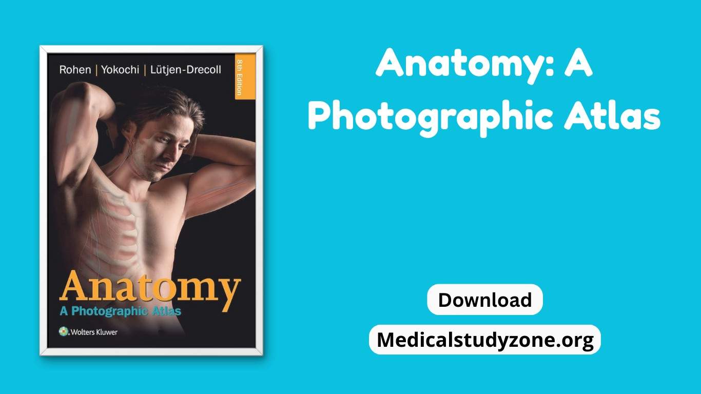
As a medical student, anatomy isn’t just about memorizing names — it’s about seeing, understanding, and being confident in the dissection room, on clinical rotations, and when interpreting scans. The right anatomy atlas can transform your study from rote learning to truly visual, contextual understanding. In this post, we’ll explore the best anatomy atlases available, and provide you this Anatomy A Photographic Atlas by Rohen PDF Free for the best of your carrier.
Standout features (what makes this atlas special)
- Huge visual library — 1209 figures, of which 1096 are in color, plus 113 radiographs, CT and MRI scans. That’s a massive photographic resource for learning by sight.
- Photo + drawing pairing — photographs of real specimens are accompanied by adapted schematic drawings so you both see and understand the structures.
- Practical organization — systematic anatomy first (bones, joints, muscles, nerves, vessels), then regional dissection chapters — ideal for pre-dissection prep and in-lab referencing.
- Imaging correlations — curated MRI/CT scans taken in planes matching specimen sections (especially useful for clinical correlations).
- Didactic clarity — the authors intentionally limited the text to essentials and focused on clear, self-explanatory presentation for students.
Top Anatomy Atlases & Tools to Consider
Here are some atlases and apps that are making waves, based on recent studies and reviews:
- Visible Body Human Anatomy Atlas — includes thousands of 3D models, micro and gross anatomy, cadaver simulations, imaging correlations. Great for interactive learning.
- VOKA Anatomy Pro — offers medically accurate 3D models, detailed pathology, augmented reality mode, and is designed to be used anywhere, anytime (both on and off-line).
- Many others apps have been evaluated via the MARS scoring system. A 2025 study found that apps like Organos internos 3D, Sistema óseo en 3D, and VOKA Anatomy Pro had some of the highest quality ratings among free anatomy apps
Book Details
| Detail | Information |
| Title | Anatomy: A Photographic Atlas PDF |
| Editor | Johannes W. Rohen, Chihiro Yokochi, Elke Lütjen-Drecoll |
| Edition | 8th Edition |
| File Size | 140 MB |
| Pages | 562 |
| Subject | Human Anatomy, Cadaveric Dissection, Medical Atlas |
| Download/Read | Available |
| Storage | Google Drive |
| Format | Free Downloadable PDF |
Download Anatomy A Photographic Atlas by Rohen PDF
Download or Read Anatomy: A Photographic Atlas Free PDF
Disclaimer: Medicalstudyzone.org does not host any files on its servers. All links are collected from publicly available sources on the internet.
We do not claim copyright for any content shared. If you believe your copyright has been infringed, please contact us for removal.
This content is provided for educational purposes only. We encourage users to purchase original licensed books and materials.
For DMCA / removal requests, email us at [email protected].

